- Original Article
- Infection
- Febrile urinary tract infection in children: changes in epidemiology, etiology, and antibiotic resistance patterns over a decade
-
Woosuck Suh, Bi Na Kim, Hyun Mi Kang, Eun Ae Yang, Jung-Woo Rhim, Kyung-Yil Lee
-
Clin Exp Pediatr. 2021;64(6):293-300. Published online October 14, 2020
-
|
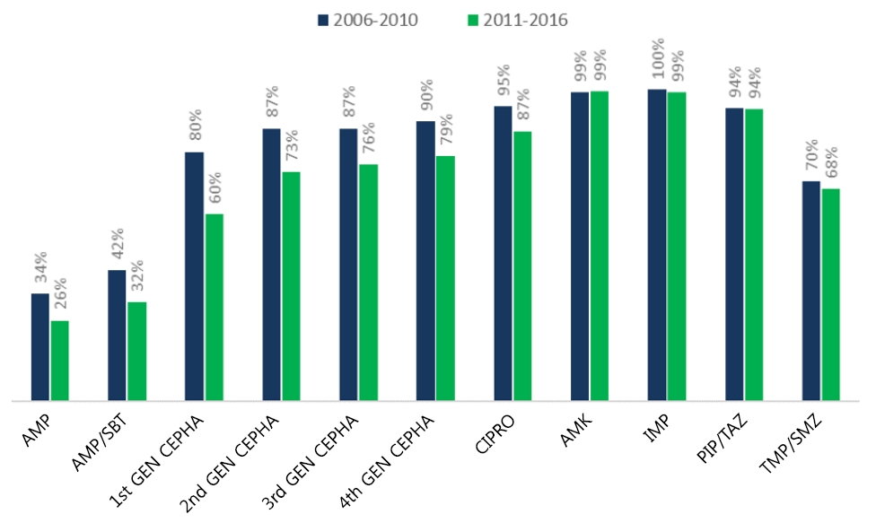
Question: How has the antibiotic susceptibility of urinary pathogens changed and what does it imply?
Finding: A yearly increase in multidrug-resistant and extended-spectrum β-lactamase (ESBL)–producing pathogens was observed. A higher recurrence rate was observed in cases of febrile urinary tract infection caused by ESBL producers in patients with underlying vesicoureteral reflux (VUR).
Meaning: The initial empirical antibiotic should reflect the changing susceptibility patterns and underlying VUR status. |
-
-
- Original Article
- Allergy
- Nasal eosinophilia and eosinophil peroxidase in children and adolescents with rhinitis
-
Yeonu Choi, Haeun Jeon, Eun Ae Yang, Jong-Seo Yoon, Hyun Hee Kim
-
Clin Exp Pediatr. 2019;62(9):353-359. Published online April 24, 2019
-
|
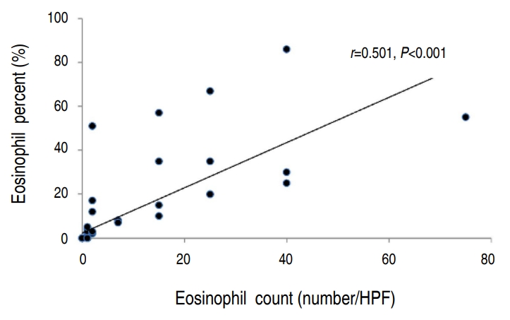
Background: Researchers have shown that eosinophil peroxidase (EPO) is a relatively accurate marker of eosinophilia and eosinophil activity. However, its use as a marker of eosinophilic inflammation in nasal secretions is limited because the diagnostic cutoff values of EPO for use as a one-time test for allergic diseases such as allergic rhinitis have not been established.
Purpose: To identify the correlation... |
-
-
- Case Report
- Secondary renal amyloidosis in a 13-year-old girl with bronchiectasis
-
Eun Ae Yang, Dong Won Lee, Myung Chul Hyun, Min Hyun Cho
-
Clin Exp Pediatr. 2010;53(7):770-773. Published online July 31, 2010
-
|
|
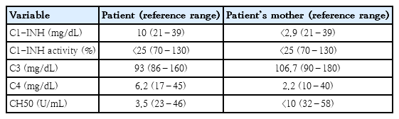
A 13-year-old girl was diagnosed with non-cystic fibrosis (CF)-related multifocal bronchiectasis accompanied by nephrotic-range proteinuria of unknown cause. On renal biopsy, there were many segmental homogeneous deposits of amyloid tissue with positive Congo red staining in the glomeruli and interstitium. On electron microscopy, relatively straight, non-branching, randomly arranged amyloid fibrils were showed in the mesangium of the glomeruli. These fibrils... |
-
-
|


")